Key Takeaways
- Emerging imaging technologies provide real-time, dynamic views of the spine.
- Noninvasive methods reduce patient radiation exposure and enhance diagnostic accuracy.
- Integration of AI and machine learning streamlines the analysis of spinal images.
Back and spine health issues are among the most common sources of discomfort for adults, creating an urgent need for safer, more effective diagnostic options. In response, medical technology has advanced rapidly over the past decade, ushering in a new era of noninvasive diagnostic solutions that promise earlier detection, greater comfort, and greater accuracy. Modern solutions, such as MRI for back pain, now allow individuals to access advanced imaging without enduring the risks often associated with invasive interventions.
Many patients experience anxiety about traditional spinal diagnostics due to concerns about radiation, discomfort, or lengthy procedures. Fortunately, innovations in noninvasive imaging have revolutionized the patient experience by offering dynamic, real-time visualizations and safer technologies tailored to diverse clinical needs. These advancements provide healthcare professionals with better data and help patients receive tailored, effective treatment plans.
Today’s noninvasive techniques don’t just improve comfort they also empower clinicians to deliver precision diagnostics, reducing unnecessary exploratory surgery and minimizing complications. With the continual integration of artificial intelligence (AI), these imaging technologies are accelerating the pace of accurate diagnosis and supporting optimal patient outcomes.
As healthcare systems adopt new diagnostic protocols, the implications reach beyond just improved imaging they touch postoperative recovery, patient education, and even the potential for remote consultations. Noninvasive imaging is no longer a luxury but an essential standard transforming modern spine care worldwide.
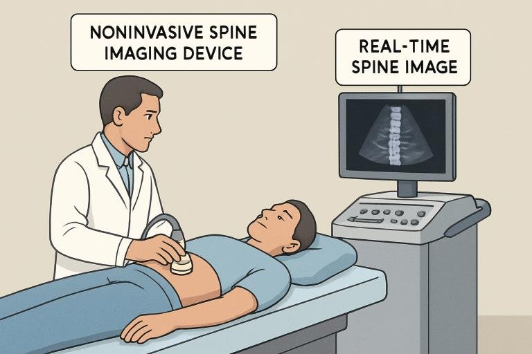
Real-Time Panoramic Ultrasound Imaging
Traditional CT and MRI techniques have long been the backbone of spinal diagnostics; however, they are inherently limited by static imaging and require patients to remain absolutely still during scanning. A breakthrough developed by researchers at Stanford Medicine involves panoramic ultrasound imaging enabled by a wide transducer and diverging-wave compounding. This approach provides an extended, dynamic view of the spine and its associated structures in real time, allowing clinicians to observe functional movements and subtle abnormalities that are missed with static imaging. This innovation improves diagnostic accuracy and may support earlier detection of complex spinal conditions. More details on this can be found at ScienceDaily.
MR Spectroscopy and AI Integration
Chronic low back pain is often multifactorial, making pinpointing its cause a major clinical obstacle. Aclarion’s NOCISCAN platform now combines MR spectroscopy a technology that evaluates biochemical changes in tissue with augmented intelligence. This enables precise localization of discogenic pain, differentiating problematic discs from their healthy counterparts. Such accuracy means physicians can create targeted, patient-specific interventions, reducing the risks of overtreatment or ineffective therapies. More on this technology and its impact on discogenic pain management is highlighted in recent Medscape coverage.
Automated MRI Segmentation with AI
Manual segmentation of spinal structures within MRI scans has historically been labor-intensive and inconsistent due to human variability. The emergence of AI-powered segmentation models has resolved many of these challenges by automating the process and achieving high reliability across different MRI systems and scan qualities. Leveraging data from vast clinical repositories and advanced neural networks, these solutions significantly expedite radiology workflows, ensuring more consistent diagnostics and timely reporting for patients and practitioners alike.
Functional Ultrasound Imaging During Surgery
During spinal surgeries, functional ultrasound imaging (fUSI) has gained traction for its ability to deliver high-resolution, real-time images of the spinal cord. Unlike preoperative scans, fUSI provides surgeons with immediate feedback during delicate procedures, allowing dynamic assessment of neural tissue vitality and function. Portability and straightforward operation have helped fUSI devices become indispensable for intraoperative spinal monitoring, potentially preventing surgical complications and improving neurological outcomes.
Minimally Invasive Endoscopic Spine Surgery
The landscape of spinal surgery has also shifted dramatically, thanks to endoscopic techniques requiring only tiny incisions. These minimally invasive approaches mean reduced post-surgical pain, less soft tissue trauma, and quicker patient recovery. A pivotal study published in World Neurosurgery demonstrated that patients undergoing unilateral biportal endoscopic discectomy used 21% fewer opioids post-surgery compared to those treated with other minimally invasive methods. Minimally invasive surgery represents a paradigm shift helping minimize complications associated with prolonged opioid use and facilitating faster reintegration into daily activities.
Integration of Robotics and AI in Spine Care
The future of spine care is defined by remarkable synergy between robotics, wearable technology, and AI-driven diagnostics. Robotic-assisted surgeries now offer greater precision for complex spinal procedures, reducing the risks of manual error. Meanwhile, wearable sensors provide real-time feedback on spinal biomechanics, supporting ongoing patient monitoring outside clinical settings. The North American Spine Society’s 2024 Annual Meeting spotlighted these disruptive technologies underscoring their value in improving overall patient safety, accelerating postoperative recovery, and increasing cost-effectiveness within spine care.
Conclusion
Spinal imaging and diagnostics are undergoing a rapid transformation as noninvasive methods become both the norm and the gold standard. Panoramic, real-time ultrasound imaging, AI-powered segmentation, advanced MR spectroscopy, and precision-guided surgery mark a significant departure from traditional practices offering patients enhanced safety, comfort, and outcomes. Keeping pace with these developments is vital for both patients seeking the most advanced care and clinicians committed to clinical excellence in spine health.